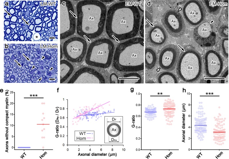
Fig. 3. Increase of thin myelin, small axons, and axons without myelin in L-MPZ mice.
a, b Representative toluidine blue staining of 500 nm-thick sections of sciatic nerves in 8-week-old wild-type (WT) and homozygous (Hom) mice. Asterisk in a indicates the lumen of blood vessels. In light micrographs (LM), compared with WT (a, arrows), compact myelin appeared thinner (b, arrows), and smaller axons with myelin ensheathment (b, arrowheads) were prominent in Hom mice. c, d In electron micrographs (EM), axons (Ax) had thick compact myelin sheath in WT (c, arrows), while axons had thin myelin sheath (d, arrows) or lacked compact myelin (d, black arrowhead) in Hom mice. Cajal bands were not clearly formed around some axons in Hom mice (d, white arrowheads). e The number of axons that were individually ensheathed by Schwann cells but lacked compact myelin (d, black arrowhead) increased in Hom mice. f–h The G-ratio (obtained by dividing axonal diameter [DAx] with fiber diameter [DF]) was larger (f, g) and axonal diameter smaller (h) in Hom mice. Bars, 10 µm (a, b) or 5 µm (c, d). **p < 0.01; ***p < 0.001 by Mann–Whitney U-test. N (photo areas prepared from each of 4 mice) in WT = 11, Hom = 11 (e). N (axons in photo areas) in WT = 115, Ho = 91 (f–h).

