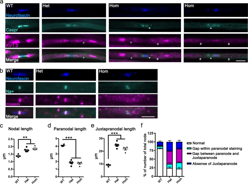
Fig. 5. Morphological abnormalities in the node of Ranvier and surrounding regions in L-MPZ mice.
a Triple immunostaining of teased sciatic nerves in 10-week-old L-MPZ wild-type (WT), heterozygous (Het), and homozygous (Hom) mice using anti-neurofascin (blue), anti-Caspr (turquoise), and anti-Kv1.2 (magenta) antibodies. White circle (○), white asterisk (*), and # indicate either gaps between the paranode and juxtaparanode, gaps within paranodal staining, or absence of Kv1.2 clusters in the juxtaparanode, respectively. Bar, 10 µm. b Triple immunostaining of teased sciatic nerves using anti-neurofascin (blue), anti-sodium channel (turquoise), and anti-moesin (magenta) antibodies. Bar, 10 µm. c–e Measurement of nodal (sodium channel cluster; c), paranodal (Caspr cluster; d), and juxtaparanodal (Kv1.2 cluster; e) lengths in the sciatic nerve of L-MPZ mice. Nodal and juxtaparanodal lengths were increased while paranodal lengths were decreased in L-MPZ Hom and Het mice. f Quantification of the number of abnormal node–paranode–juxtaparanode structures in sciatic nerve sections of L-MPZ mice. Abnormal structures were categorized into three (turquoise column, white asterisk; magenta column, white circles; blue column, white #) as described in a. The percentage of nodes with abnormal node–paranode–juxtaparanode structures was increased in L-MPZ mice. **p < 0.01; ***p < 0.001 by one-way ANOVA with post-hoc Tukey’s test. Data are presented as mean ± SE of experiments. N (mice) in WT = 4, Het = 6, Hom = 4 (c–f).

