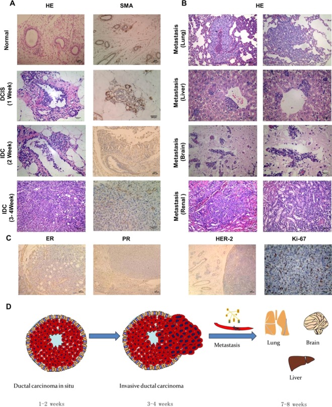
Figure 3.
Pathological features of tumor and visceral metastases in the 4T1-MIND model. (A) Representative photographs of H&E staining, SMA immunohistochemical staining sections of normal ducts and the mammary glands of female BALB/C mice injected with 4T1 cells. The ductal architecture demarcated by SMA in the normal breast ducts (n = 5). In the first week, a DCIS-like structure formed in the female BALB/C mice injected with 4T1 cells (n = 5), SMA immunostained the myoepithelial layer around the DCIS. In the second week, Tumor cells have just broken through the breast duct and grow outside of them, SMA immunostained the Part of MECs around the breast ducts (n = 5). IDC-like structure formed in the female BALB/C mice injected with 4T1 cells for 3–4 weeks (n = 5), the MECs almost disappears and expression of SMA was negative. Scale bar, 100–200 μm. (B) Representative photographs of H&E staining sections of the viscera of female BALB/C mice injected with 4T1 cells for 7 weeks (n = 5). Lung, brain and renal metastasis could be observed. Tumor cell metastasis from the central vein forms liver metastasis. Scale bar, 100–200μm. (C) Representative immunohistochemical staining sections of the tumors of female BALB/C mice injected with 4T1 cells for 4 weeks (n = 5). The ER, PR and HER-2 immunostaining sections of transplanted tumor were negative (positive expression around normal mammary duct), but the expression of Ki-67 was positive. Scale bar, 100–200 μm. (D) Schematic diagram of model development.

