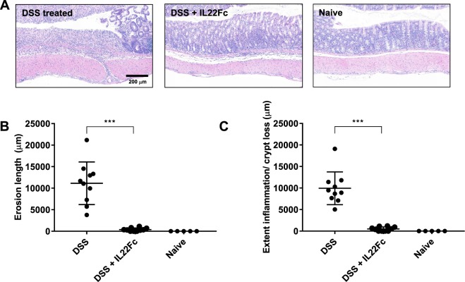
Figure 5.
Histological assessment of epithelial erosions in C57BL/6 mouse colon. (A) Characteristic H&E stains: left: ulceration of the epithelial layer, crypt damage, and infiltration of inflammatory cells into the colon mucosa in DSS treated animals; middle: reduction in epithelial damage for DSS + IL22Fc treated animals; right: naive animal. (B,C) Erosion length, extent of inflammation, and crypt loss analyses suggest DSS + IL22Fc treatment provides epithelial protection at near naive levels. For DSS, n = 10; for DSS + IL22Fc, n = 10; for naive, n = 5. All values presented are the mean ± S.D. One-way ANOVA was used to analyze variation; ***P < 0.0001.

