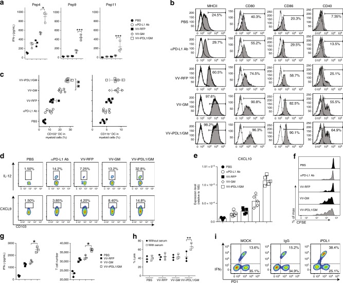
Fig. 7. Enhanced neoantigen presentation and cytolytic activity of neoantigen-specific CTLs.
a Enhanced stimulatory potency of tumor-infiltrating DCs. Tumor-infiltrating DCs from VV-treated mice were loaded with neopeptide 4, 9, or 11, and cocultured with the neoantigens-primed T cells from mice immunized with the 11 neopeptide mixture to assess IFN-γ production; n = 3 mice. Data presented as the means ± SD. *P < 0.05, ***P < 0.001 by two-tailed Student’s t-test. b Enhanced maturation of tumor-infiltrating DCs. Using a similar treatment schedule as described in Fig. 5a, cell suspensions prepared from VV-treated tumors were analyzed by flow cytometry. c Enhanced tumor infiltration of CD103+ DCs. Using the same treatment schedule as in Fig. 5a, tumor cell suspensions from VV-treated mice were analyzed by FACS; n = 5 mice. Data presented as the means ± SD. **P < 0.01 by two-tailed Student’s t-test. d Intracellular staining of IL-12 and CXCL9 of CD103+ DCs from VV-treated tumors. e qRT-PCR analysis of CXCL10 mRNA levels in CD103+ DCs isolated from VV-treated tumors; n = 5 mice. Data presented as the means ± SD. **P < 0.01 by two-tailed Student’s t-test. f Neoantigens-primed T cells proliferated more efficiently in VV-iPDL1/GM-treated mice. The neoantigens-primed T cells were labeled with 5 μM CFSE and i.v. injected into VV-treated mice. Three days later, T cell proliferation was assessed by FACS. g Enhanced stimulatory effect of VV-iPDL1/GM-infected tumor cells. MC38 tumor cells infected with VVs at MOI = 1 were cocultured with the neoantigens-primed T cells. IFN-γ production (left) and T cell proliferation (right) were measured. Data presented as the means ± SD. *P < 0.05 by two-tailed Student’s t-test. h Serum of VV-iPDL1/GM-treated mice enhanced the cytolytic activity of neoantigens-primed T cells. MC38-Luc cells were cocultured with the neoantigen-specific T cells in the presence of the sera from treated MC38-bearing mice. Cytolytic activity was calculated using luciferase emission value. Data are presented as means ± SD. **P < 0.01 by two-tailed Student’s t-test. i PD-1+ CD8+ T cells isolated from VV-treated MC38 tumors were cocultured with MC38 cells in the presence of purified iPDL1 or IgG. IFN-γ+ frequencies of PD-1+ T cells were shown from one of two independent experiments.

