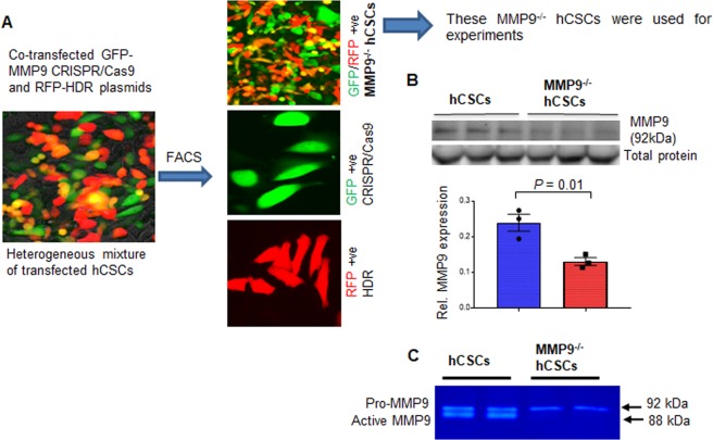
Fig. 3. Creation and validation of MMP9−/− hCSCs.
CRISPR-Cas9 method was used to generate MMP9−/− hCSCs. a Representative image showing heterogeneous hCSCs after co-transfection with CRISPR-Cas9 (green) and HDR (red) plasmids. Representative images of three groups of hCSCs after fluorescence-activated cell sorting (FACS). b Western blotting and densitometric qualification of MMP9 protein validating decreased MMP9 expression in MMP9 ablated hCSCs (MMP9−/− hCSCs). β-actin is a loading control. Each dot represents one sample. Values are expressed as mean ± SEM. Student t-test was used. P < 0.05 is considered statistically significant. c Representative in-gel-gelatin zymography showing decreased MMP9 activity in MMP9−/− hCSCs.

