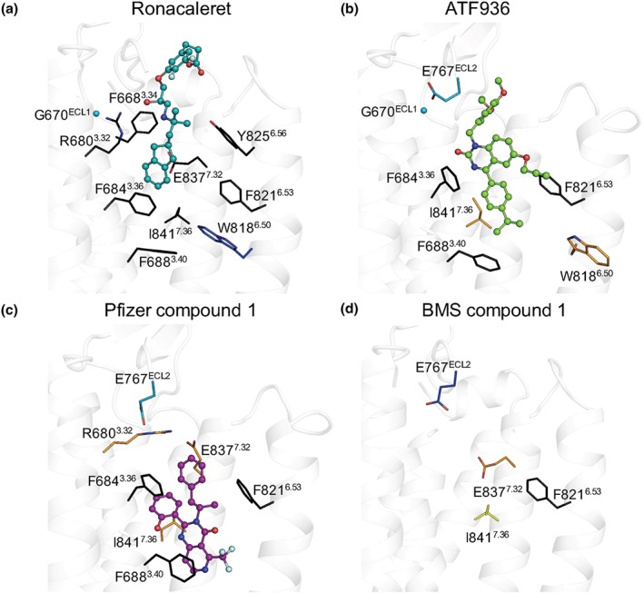
Figure 4.

Mutagenesis and computational modelling predict CaS receptor NAMs bind in overlapping but disparate binding orientations within the 7TM domain. (a) Ronacaleret, (b) ATF936, and (c) Pfizer compound 1 are shown in sticks and balls docked in the 7TM domain (white helical ribbons). Mutated residues are shown as sticks and are coloured according to a greater than twofold increase (light teal), greater than fivefold increase (dark blue), greater than twofold decrease (yellow), or greater than fivefold decrease (orange) in binding affinity. When affinity could not be determined (i.e. a complete abolishment of NAM activity), residues are shown in black. Salt bridges are shown as red dashes, and Gly670ECL1 is shown as a sphere. While residues that contribute to BMS compound 1 affinity are shown (d), BMS compound 1 is not docked in the 7TM because predicted docking poses were not compatible with the mutagenesis or SAR data
