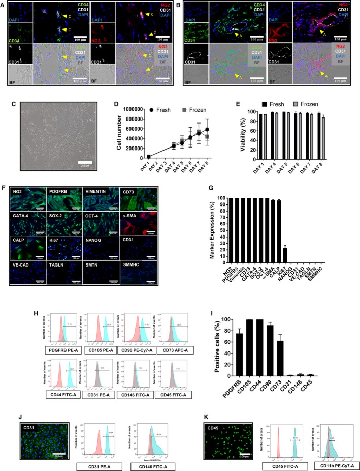
Figure 1.

Characterization of swine cardiac pericytes. (A and B) Perivascular localization of sCPs in situ. Immunofluorescence images showing the localization of CD31–/CD34+/NG2+ CPs in swine hearts around capillaries (A) and arterioles (B). Inserts showing CD34 labeled in green fluorescence, NG2 in red, and CD31 in white; nuclei are recognized by blue fluorescence of DAPI. Arrows indicate CPs around capillaries and an arteriole. Images taken at 200× magnification. C, Phase contrast microscopy image of sCPs displaying spindle‐shape features (magnification, ×100). (D and E) Graphs showing the growth curve (D) and viability (E) of 3 sCP lines that were seeded at 3000 cells/cm2 at day 1 and detached and counted at days 4, 5, 6, 7, and 8 of culture. F, Immunofluorescence microphotographs showing the expression of neural/glial antigen 2 (NG2) and platelet‐derived growth factor receptor beta (PDGFRB), vimentin, CD73, cardiac transcriptional factor GATA‐binding protein 4 (GATA‐4), and the stemness markers, sex determining region Y‐box 2 (SOX‐2) and octamer‐binding transcription factor 4 (OCT‐4). Cells are negative for NANOG and the endothelial cell markers, vascular endothelial‐cadherin (VE‐cadherin) and CD31. sCPs express alpha‐smooth muscle actin (α‐SMA) and calponin (CALP) and are negative for transgelin (TAGLN), smoothelin (SMTN), and smooth muscle myosin heavy chain (SMMHC). Expression of Ki67 is indicative of proliferating cells. DAPI (blue) identifies nuclei. Scale bars=100 μm. G, Values in bar graph represent the mean±SEM of 7 biological replicates from immunocytochemistry studies. Fluorescence was normalized by nuclei count. (H and I) Flow cytometry analysis of 3 sCP lines at P5. H, Representative graphs for each surface marker; negative control staining profiles are shown by the red histograms, whereas specific antibody staining profiles are shown by light blue histograms. I, Bar graph shows the mean±SEM values of 3 sCP lines. (J and K) Immunofluorescent images and flow cytometry histograms are also shown for swine pulmonary artery endothelial cells (PAECs) (J) and peripheral blood mononuclear cells (PBMNCs; K). APC indicates allophycocyanin; BF, bright field; CD, cluster of differentiation; DAPI, 4′,6‐diamidin‐2‐fenilindolo; FITC‐A, fluorescein‐area; PE‐A, phycoerythrin‐area; PE‐Cy7, phycoerythrin–cyanine 7; sCP, swine cardiac pericytes.
