
Figure 3.
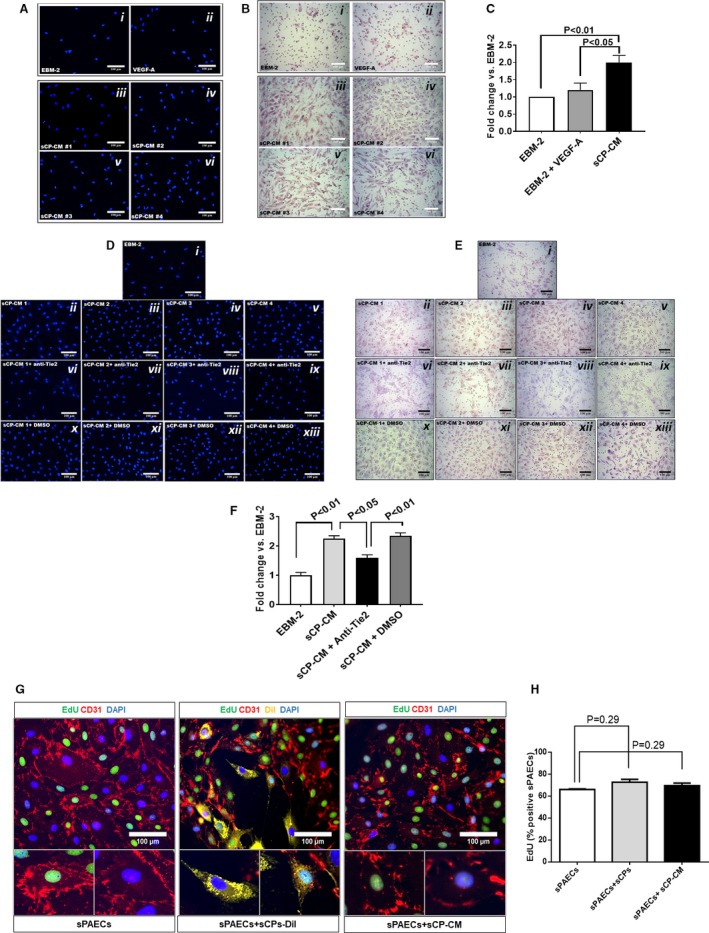
Chemotactic activity of factors secreted by swine cardiac pericytes. (A through C) In a transwell migration assay, sCP‐CM enhanced the migration of sPAECs. Representative images of migrated cells stained by DAPI (A) or Giemsa (B) following stimulation with EBM‐2 (i), VEGF‐A (ii, 100 ng/mL), or CM from 4 sCP lines (iii–vi). Images acquired using a 200× magnification. (C) Bar graph showing the fold change of migrated cells vs EBM‐2 basal media. N=4, data are mean±SEM. (D through F) Effect of Tie‐2 inhibitor (7.5 μmol/L) on the chemotactic activity of sCP‐CM. Representative images of migrated cells stained by DAPI (D), or Giemsa (E) following stimulation with EBM‐2 (i), CM from 4 sCP lines (ii–v), or CM from the same sCP lines added with a Tie‐2 antagonist (vi–ix) or its vehicle (x–xiii, DMSO). Images acquired using a 200× magnification. F, Bar graph showing the fold change of migrated cells vs EBM‐2 basal media. Data are mean±SEM. Representative immunofluorescent images of proliferating sPAECs following coculture with sCPs and sCP‐CM. (G) sPAECs are stained with anti‐CD31 (red fluorescence) and sCPs with the long‐term cell tracker, Dil (yellow fluorescence). Images acquired using 200× magnification. (H) Bar graph displaying the percentage of EdU+ sPAECS following stimulation with sCPs and sCP‐CM. N=4, data are mean±SEM. CD indicates cluster of differentiation; Dil, chloromethylbenzamido; DMSO, dimethyl sulfoxide; EBM‐2, endothelial cell basal medium‐2; EdU, 5‐ethynyl‐2′‐deoxyuridine; sCP‐CM, swine cardiac pericyte–conditioned media; sPAECs, swine pulmonary artery endothelial cells; Tie2, tyrosine kinase 2; VEGF‐A, vascular endothelial growth factor A.