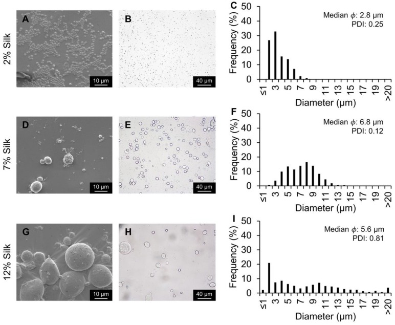
Figure 5.
Silk particles fabricated using the 20 µm flow-focusing microfluidics device and varying the dispersed phase silk concentration. The dispersed phase concentration was either 2%, 7%, or 12% silk at 0.8 mL/h. The external phase concentration was 5% PVA. (A,D,G) SEM and (B,E,H) brightfield microscopy images of silk particles. (C,F,I) Silk particle size distribution, median, and PDI measured via image analysis.

