Figure 2.
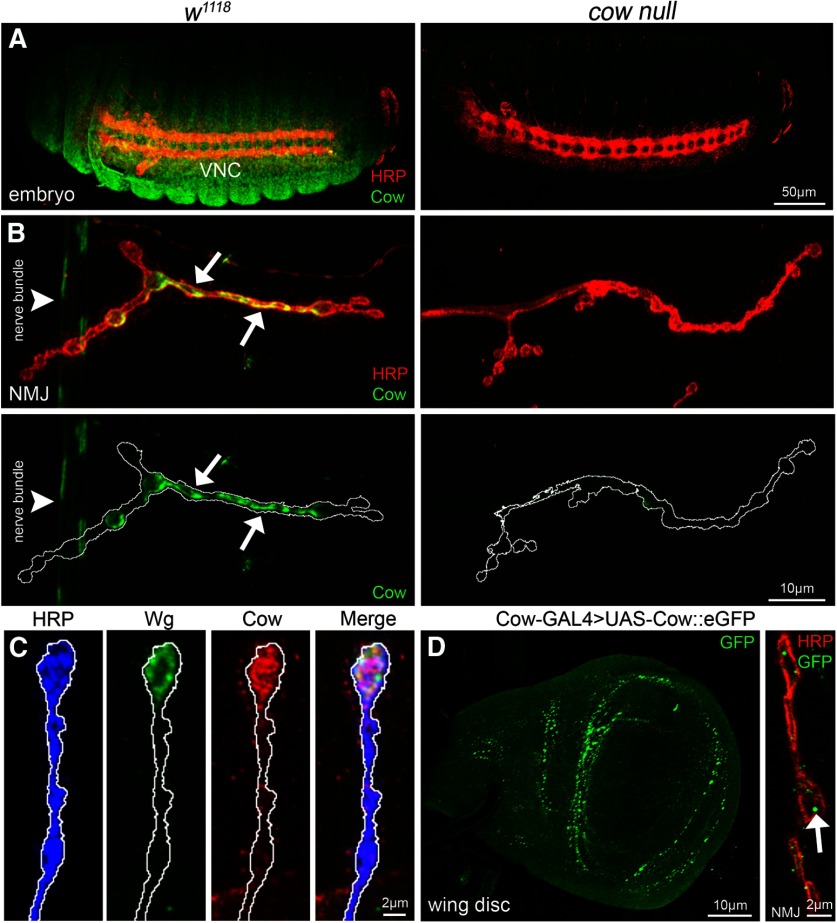
Cow expression in embryos, larval NMJ synaptic terminal, and wing disk. A, Confocal images of stage 16 embryos colabeled with anti-HRP (red) to mark neuronal membranes and anti-Cow (green) in genetic background control (w1118, left) and cow null (cowGDP/cowGDP, right). The ventral nerve cord (VNC) is labeled. B, Confocal images of third instar NMJ colabeled with anti-HRP (red) and anti-Cow (green) in control (w1118, left) and cow null (cowGDP/cowGDP, right). From nonpermeabilized labeling, Cow appears secreted from a dynamic subset of synaptic boutons (arrows) and also present in the nerve bundle (arrowhead). Cow is shown without HRP in below images. White line marks the NMJ terminal HRP domain. C, Higher-magnification images of w1118NMJ synaptic boutons colabeled with anti-HRP (blue), anti-Wg (green), and anti-Cow (red), with merged image on right. White line marks the NMJ terminal HRP domain. D, Cow-GAL4 driving UAS-Cow::eGFP in wandering third instar wing imaginal disk (left) and NMJ colabeled with anti-HRP (red) and anti-GFP (green, right). For the NMJ, a single confocal section (0.5 μm) shows Cow punctae (arrow) within and surrounding synaptic boutons.

