
Figure 4.
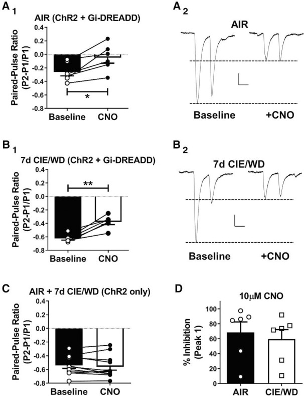
Bath application of CNO decreases glutamate release from dmPFC-BLA terminals expressing the hM4Di-DREADD receptor. A1, B1, Light-evoked paired-pulse ratios recorded from dmPFC-BLA terminals expressing ChR2 and Gi-coupled DREADDs at baseline (closed bars, open circles) and during the bath application of CNO (10 μm; open bars, closed circles) in air-exposed control neurons (N = 6; A1) and in 7 d CIE/WD neurons (N = 6; B1). Note that some individual data points are superimposed due to the scale. CNO significantly decreased the oEPSC amplitude and increased the PPR in all of the cells. *p < 0.05; **p < 0.01; paired t test. A2, B2, Representative traces of paired-pulse responses recorded from AIR (A2) or CIE/WD (B2) neurons at baseline (left) and in the presence on CNO (right). Calibration: x = 50 ms, y = 20 pA. C, PPRs recorded from AIR and CIE/WD BLA neurons (N = 11) with dmPFC-BLA terminals expressing only ChR2 (no DREADD controls). CNO did not significantly change glutamate the release probability from terminals without expressing the DREADD construct. D, The percentage of inhibition by CNO, calculated from the first response amplitude (A2, B2), was not different between AIR and CIE/WD recordings.