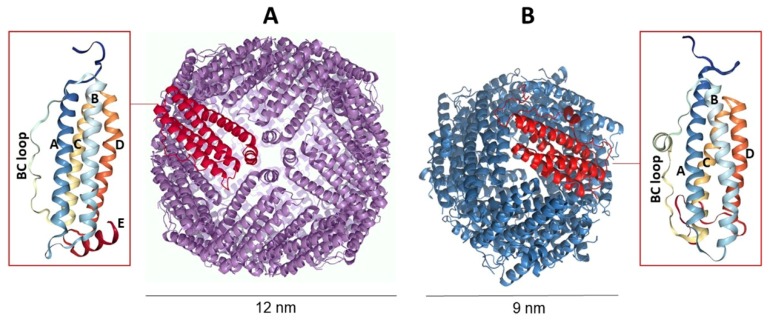
Figure 1.
Comparison between canonical ferritin (A) and DNA-binding protein from starved cells (Dps) (B). While canonical ferritins form a cage of 24 subunits arranged in octahedral 4-3-2 symmetry, Dps proteins are dodecamers displaying 2- and 3-symmetry axes. The insets show the monomer structure consisting of a bundle of α-helices marked with capital letters.

