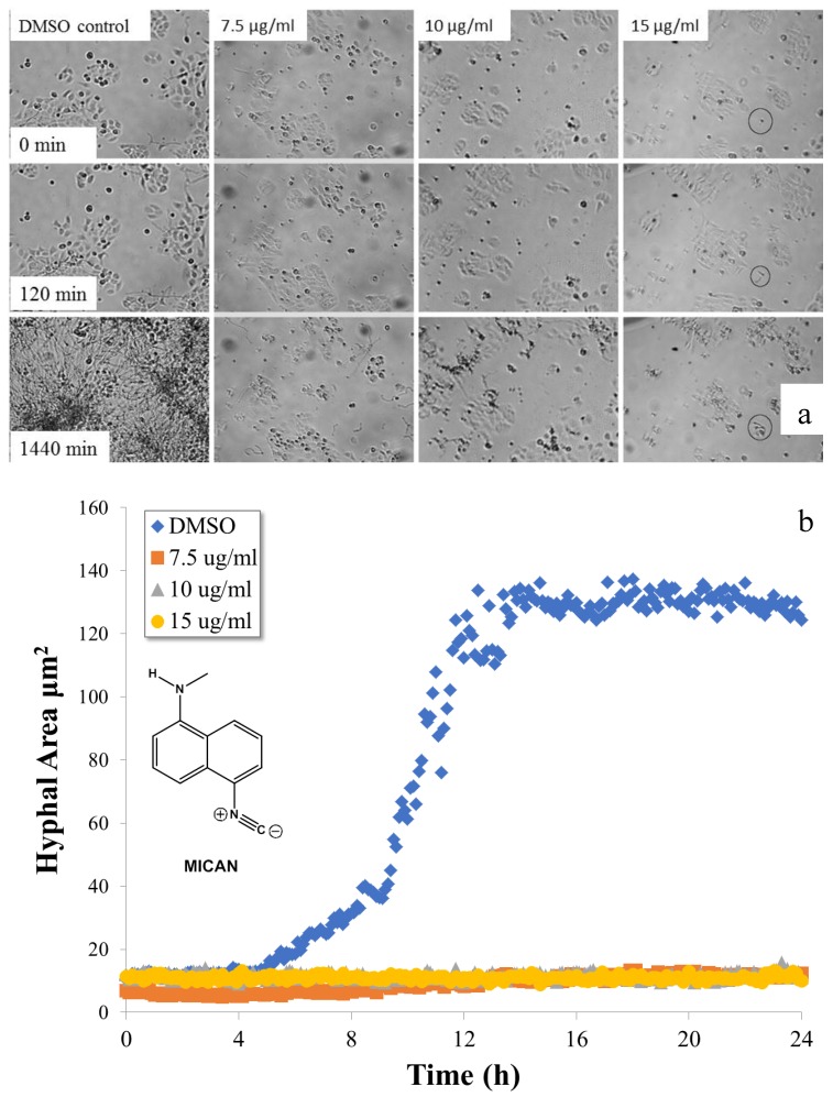
Figure 1.
Hyphal growth of C. albicans in the presence of methylamino-5-isocyanonaphthalene (MICAN). Time-lapse microscopic images (a) of HaCat cells infected by C. albicans in the presence of different concentrations of MICAN and the corresponding hyphal growth curves (b) determined from the average individual hyphal area of C. albicans. (Areas were measured instead of lengths due to the hyphal ramifications).

