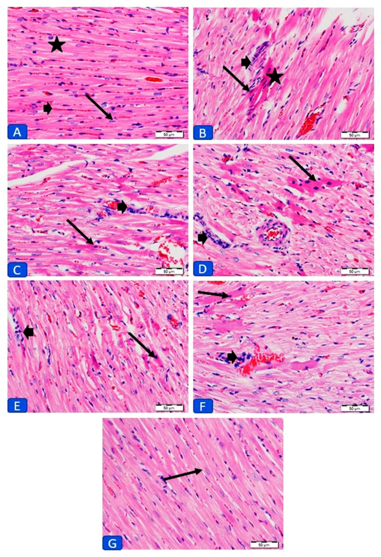
Figure 2.
Photomicrographs of sections in the left ventricles (LV) of (A) control rats showing normal histology of cardiomyocyte cytoplasm (star) and nuclei (arrows) and normal distribution of endomysium (bold arrow). (B) DOX-intoxicated rats showing foci of degenerated cardiomyocytes and collection of inflammatory cells in the endomysium (bold arrow). (C) CAR-treated rats showing mild improvement of myocardium with less degeneration (arrow) and inflammatory cells (bold arrow). (D) RES-treated rats showing foci of degenerated cardiomyocytes (arrow) and few inflammatory cells (bold arrow). (E) CAR/RES- and (F) LIPO-RES-treated rats showing moderate decrease in degeneration (arrow) and inflammatory cells (bold arrow); and (G) CAR/LIPO-RES-treated rats with no degeneration or inflammatory cells and the cardiomyocytes appear normal. (× 400; H&E; scale bar: 50 µm).

