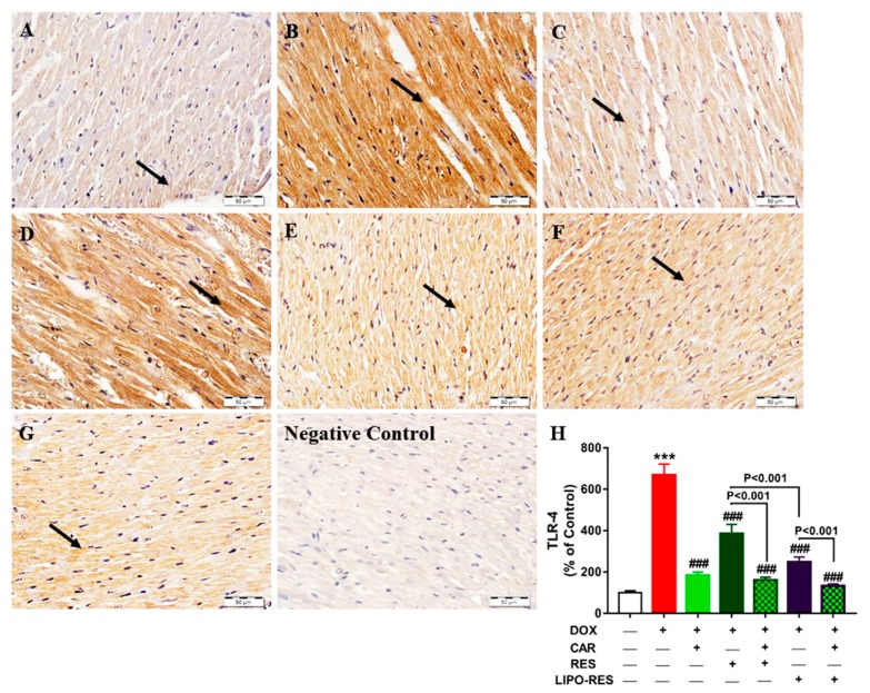
Figure 3.
Photomicrographs of anti-TLR-4-stained LV sections in: (A) Control rats showing the absence of immune reaction in cardiomyocytes cytoplasm and nuclei (arrow); (B) DOX-intoxicated rats showing very strong immune positivity; (C) CAR-treated rats showing mild immune positive reaction; (D) RES-treated rats showing strong positive immune reaction; (E) CAR/RES-treated rats showing very mild immunostaining; (F) LIPO-RES-treated rats showing moderate immune reaction and (G) CAR/LIPO-RES-treated rats showing marked decrease in TLR-4 immune reactivity. (H) Mean ± SEM of TLR-4 immunostaining in LV sections of different groups, (n = 6). *** p < 0.001 versus Control and ### p < 0.001 versus DOX.

