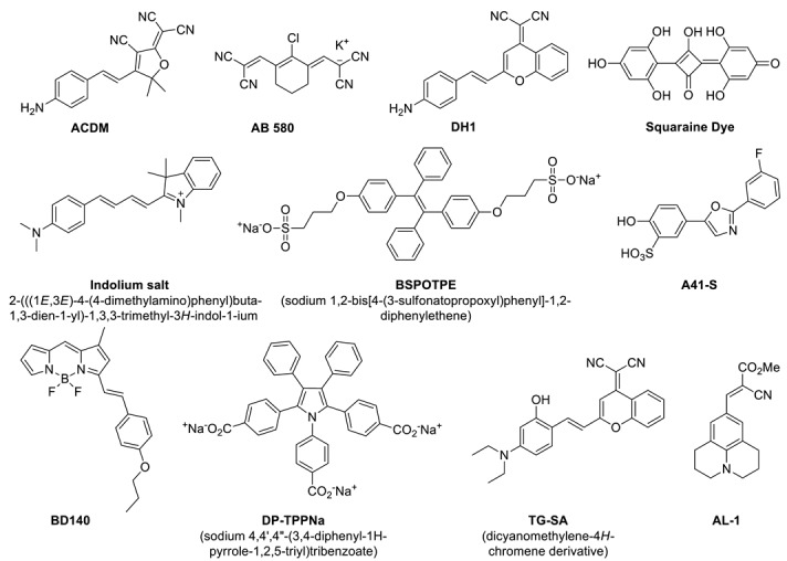
Table 1.
Fluorescent HSA sensors: structures, fluorescence properties, HSA sensitivity (fluorescence fold increase), binding sites and sensing properties (limit of detection, detection range).

| Name | Fluorescence properties | Sensing properties (mg/L) | Binding site d | Ref | |||
|---|---|---|---|---|---|---|---|
| λex (nm) a | λem (nm) b | Fold increase c | Limit of Detection | Detection range | |||
| TG-SA | 530 | 733 | 100 | 1.26 | 1.26–232 | I | 14 |
| ACDM | 560 | 612 | 75 | 2.5 | 0–300 | ND e | 15 |
| Indolium salt | 550 | 680 | 12 | 0.73 | 0.73–998 | I | 16 |
| AB 580 | 590 | 616 | 17 | 0.4 | 1–50 | NA f | 17 |
| DP-TPPNa | 310 | 443 | 9 | 1.68 | 1.68–100 | NA f | 18 |
| BSPOTPE | 350 | 475 | 300 | 0.67 | 0–6.7 | I/II f | 19 |
| A41-S | 360 | 473 | 55 | NA f | NA f | I | 20 |
| Squaraine Dye | 560 | 620 | 80 | NA f | NA f | II | 21 |
| BD140 | 520 | 585 | 41 | NA f | NA f | II | 22 |
| AL-1 | 456 | 490 | 400 | 0.4 | 0–66.5 | I | 23 |
| DH1 | 520 | 620 | 70 | 0.022 | 0-11.9 | I | 24 |
a Absorption wavelength; b Emission wavelength; c Fold increase in fluorescence intensity of the sensor after addition of HSA; d Drug binding site 1 (I) and 2 (II); e Not determined; f Not available.
