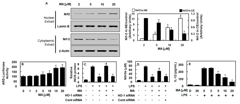
Figure 3.
MA induced the nuclear translocation of Nrf2 and anti-inflammatory action in HUVECs. (A) HUVECs were harvested, and cytosolic and nuclear fractions were extracted (CE and NE, respectively) after treatment with MA (2–20 μM) for 6 h. Western blotting was employed with indicated antibodies (Left panel), (Uncropped pictures of the Western blot (upper panel and lower panel) was shown in Figures S2 and S3, respectively), and the densitometric intensity of Nrf2 normalized to Lamin B or β-actin is shown (Right panel). (B) ARE luciferase reporter activity was measured with lysates from cells transfected with ARE. (C,D) HO-1 expression was suppressed with siRNA to determine whether MA-mediated HO-1 expression was responsible for iNOS (C) and NO (D) inhibition. (E) IL-1β concentrations were measured with an ELISA kit. The results represent the mean value with SD from three independent experiments conducted in triplicates on three different days. D denotes 0.2% DMSO treatment, which was used as the vehicle control. * p < 0.05 versus LPS, # p < 0.05 versus LPS + MA, or + p < 0.05 versus LPS + MA + HO-1 siRNA.

