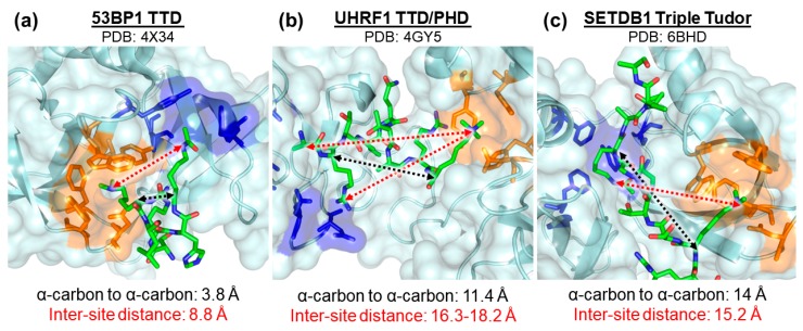
Figure 2.
Close-up view showing basic dimensions and spatial features for multivalent Kme reader: peptide interactions of proteins (a) 53BP1 (PDB: 4X34), (b) UHRF1 (PDB: 4GY5), and (c) SETDB1 (PDB: 6BHD). The regions highlighted in orange of each panel are the methyl-lysine binding pockets and the regions highlighted in blue are the ancillary binding pockets. The distances between the α-carbon of each PTM residue and the inter-site distances are depicted using black and red arrows, respectively.

