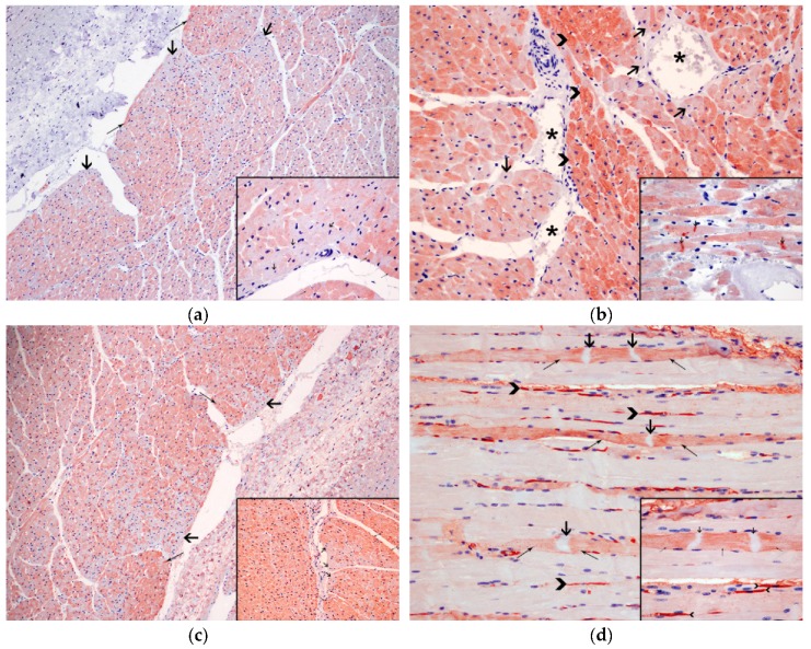
Figure 4.
Immunohistochemical techniques: (a) Degenerated cardiomyocytes (thick arrows) present depletion of cardiac troponin I (cTnI), compared to the normal cardiomyocytes (arrow heads). 10×. Inset: Detail of the intrafibrillar depletion (thick arrows) of cTnI in the injured cardiomyocytes, in comparison with normal cardiomyocytes (thin arrows); 60×, anti-troponin I; (b) Necrotic cardiomyocytes (thick arrows), close to the blood vessels (*) demonstrate depletion of cardiac troponin C (cTnC), compared to the normal cardiomyocytes (arrow heads); 20×. Inset: Detail of intrafibrillar depletion of cTnC with intense immunolabelling in the contraction band necrosis (thick arrows); 60×, anti-troponin C; (c) Degenerated cardiomyocytes (thick arrows), show intrafibrillar depletion of myoglobin, compared to the normal cardiomyocytes (thin arrows); 10×. Inset: Detail of the intrafibrillar depletion (thick arrows) of myoglobin in the injured cardiomyocytes, in comparison with normal cardiomyocytes (thin arrows); 20×, anti-myoglobin; (d) Expression of fibrinogen, in the necrotic myocytes, mainly in area next to (thin arrows) the contraction band necrosis (thick arrows). Presence of fibrinogen in the blood vessels (arrow head); 20×. Inset: Detail of the immunolabelling of fibrinogen, in the injured myocytes, in the zone near to (thin arrows) the contraction band necrosis (thick arrows). Fibrinogen inside the blood vessels (arrow heads); 60× anti-fibrinogen.

