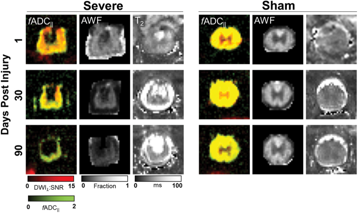
FIG. 1.
Representative MRIs. Comparison of a severe-injured rat (left) with a sham-injured rat (right) shows the evolution of cord degeneration over time after a contusion injury as measured with multiple imaging modalities. fADC|| images derived from DDE demonstrate color-coded estimates of axonal density (green) and axonal injury (red), with more axonal injury observed acutely followed by degeneration of these injured axons over time. Axonal water fraction (AWF) showed an overall decrease over time after injury and reduction compared to sham animals. Finally, T2-weighted images showed areas of increased signal in the cord with the development of fluid collections in areas of axonal degeneration. fADC|| “filtered” parallel diffusivity from imaged-based DDE; DDE, double-diffusion encoding; SNR, signal-to-noise ratio. Color image is available online.

