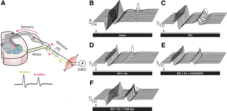
FIG. 2.
Representative recordings of H-reflexes evoked by a train of stimulation to the tibial nerve in the interosseus muscle. (A) The stimulation of the tibial nerve evokes a volley in Ia afferents (solid pink arrows) that monosynaptically excite alpha motoneurons. The M-wave (green arrow) precedes the H-reflex (dotted pink arrow) and is due to the direct activation of motor axons. (B–F) Typical EMG recordings over a series of 20 stimulations to the tibial nerve illustrating that during a 10-Hz stimulation train, the depression of the H-reflex is impaired after SCI (C) as compared with intacts (B) but is substantially restored in exercised animals (D). However, the exercised groups that received VU0240551 (E) or TrkB-IgG (F) during the daily rehabilitation session exhibited a very modest depression as compared with exercised animals. Overall, blocking KCC2 or BDNF activity in exercised animals (E–F) yields responses similar to non-exercised SCI (C). EMG, electromyogram; SCI, spinal cord injury.

