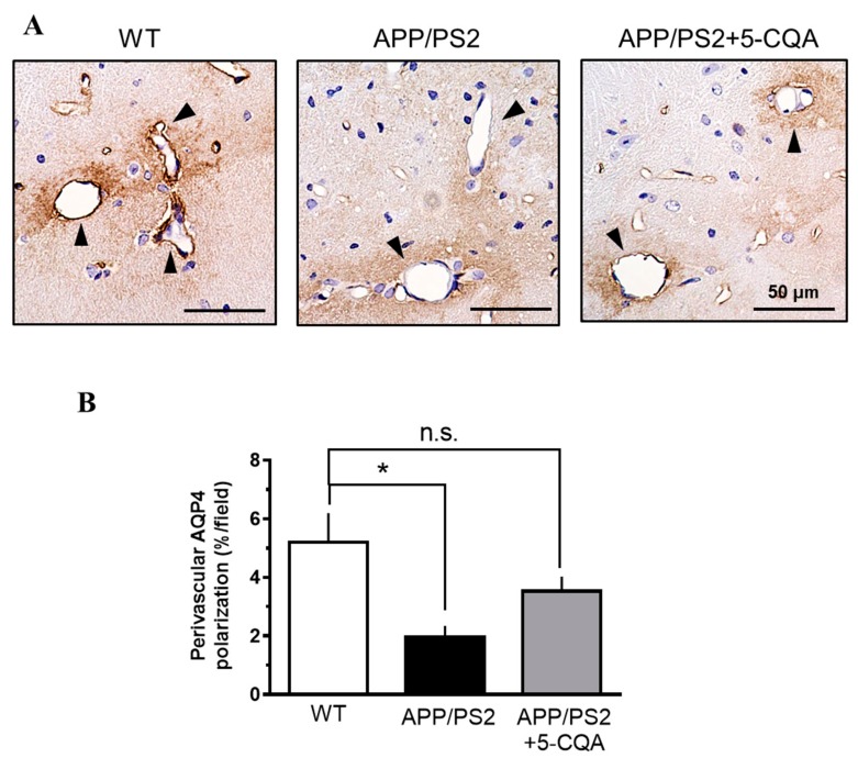
Figure 6.
Perivascular AQP4 polarization in APP/PS2 mouse hippocampus. (A) Representative AQP4 immunoreactive images of hippocampus in WT, APP/PS2, and APP/PS2 + 5-CQA mice. AQP4 expression closely abutting vessels (black arrows). Scale bar: 50 µm at ×40 magnification. (B) Quantification of perivascular AQP4 polarization. Data are means ± SEM (n = 6–8 mice/treatment). *: p < 0.05 vs. APP/PS2 (Bonferroni’s post-hoc test).

