Figure 1.
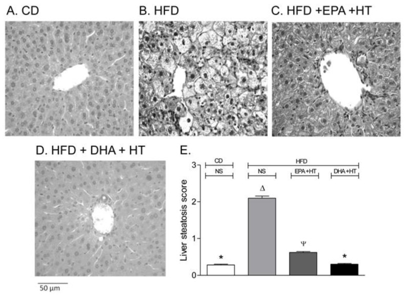
Liver Histological assessment in mice subjected to (A) control diet (CD), (B) high-fat diet (HFD) without supplementation (NS), (C) HFD supplemented with eicosapentaenoic acid (EPA) plus hydroxytyrosol (HT) and (D) HFD, supplemented with docosahexaenoic acid (DHA) plus HT. Weaning male C57BL/6J mice (n = 7 per experimental group) were allowed free access to a CD (10% fat, 20% protein, and 70% carbohydrate, with a caloric values of 3.85 KcaL/g; Rodent Diet, Product data D12450B and D12492, Research Diet Inc., USA) or HFD (60% fat, 20% protein, and 20% carbohydrate, with a caloric values of 5.24 Kcal/g; Rodent Diet, Product data D12492, Research Diet Inc., USA) for 12 weeks. Animals subjected to CD (not shown) or HFD were simultaneously supplemented with EPA (50 mg/kg/day) plus HT (5 mg/kg/day) or DHA (50 mg/kg/day) plus HT (5 mg/kg/day) through gavage. Liver samples were fixed in phosphate-buffered formalin, embedded in paraffin, stained with haematoxylin-eosin, and analyzed by optical microscopy in blind fashion describing the presence of steatosis, graded according to Brunt et al. [32]. (E) Liver steatosis scores (mean ± SEM; n = 7). a,b,c,d Groups sharing the same lyrics are not significantly different among them according to a two‐way ANOVA and Bonferroni’s post-test (p < 0.05). Adapted from Echeverría et al. [38] and Soto-Alarcón et al. [56].

