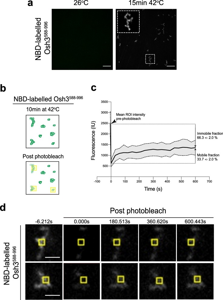
Fig. 7.
The Osh3 PI4P-binding ORD region forms condensates in vitro. a Purified NBD-labeled Osh3588–996 was imaged at 26 °C and after incubation at 42 °C. Upon elevated temperature, NBD-labeled Osh3588–996 assembles into visible condensates. The inset shows a × 5 magnification of an example of an Osh3588–996 condensate. Scale bars, 10 μm. b Scheme displaying fluorescence recovery after photobleaching (FRAP) analyses on NBD-labeled Osh3588–996 condensates formed after a 15-min incubation at 42 °C. The yellow boxes mark the regions subjected to five sequential 200 ms rounds of photobleaching. Fluorescence intensity decreased throughout the condensates upon photobleaching (see Fig. 6d for examples). c Quantitative FRAP analyses of NBD-labeled Osh3588–996 condensates formed after incubation at 42 °C. The mean intensity of regions of interest (ROI) from 8 condensates from two independent experiments is shown prior to (arrowhead) and following photobleaching (solid black line). Standard deviation is shown (gray area between dashed lines). The mobile and immobile fractions are indicated. d NBD-labeled Osh3588–996 condensates formed after incubation at 42 °C were placed on a coverslip for time-lapse FRAP imaging. Images were taken at the time points indicated. Images show individual NBD-labeled Osh3588–996 condensates and the yellow partitions demarcate the photobleached regions of interest (ROI). Scale bars, 2 μm

