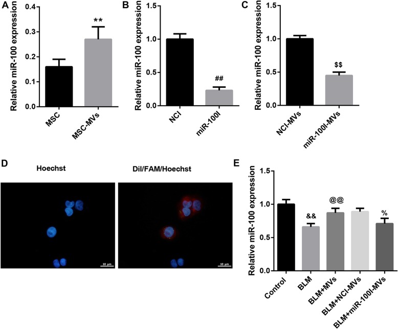
Fig. 1.
MSC-MVs mediate transfer of miR-100 to BLM-treated L2 cells. a qRT-PCR analysis of miR-100 expression in MSCs and MSC-MVs. b qRT-PCR analysis of miR-100 expression in NCI-transfected MSCs and miR-100I-transfected MSCs. c qRT-PCR analysis of miR-100 expression in MVs derived from NCI-transfected MSCs (NCI-MVs) and MVs from miR-100I-transfected MSCs (miR-100I-MVs). d The MVs derived from FAM (green)-miR-100-transfected MSCs were labeled with Dil (red) and co-cultured with rat type II alveolar epithelial cell line L2 (nuclei stained with Hoechst 33342, blue). The distribution and intensity of fluorescence were observed by confocal laser microscopy to analyze MV uptake by L2 cells. e qRT-PCR analysis of miR-100 expression in L2 cells in the groups of control, BLM, BLM+MVs, BLM+NCI-MVs, and BLM+miR-100I-MVs. Data are presented as the means ± SD (n = 3). **p < 0.01, vs. MSC; ##p < 0.01, vs. NCI; $$p < 0.01, vs. NCI-MVs; &&p < 0.01, vs. control; @@p < 0.01, vs. BLM; %p < 0.05, vs. BLM+NCI-MVs

