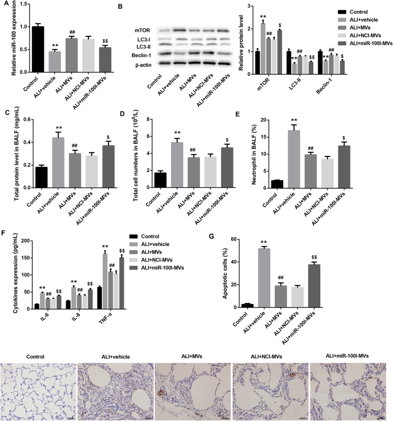
Fig. 5.
Changes of miR-100 expression, autophagy level, lung apoptosis, and inflammation in ALI rats 48 h following administration with miR-100I-MVs. qRT-PCR analysis of miR-100 in lung tissues (a); western blot analysis of protein levels of mTOR, LC3-I, LC3-II, and Beclin-1 in lung tissues (b); total protein level in BALF (c); total cell numbers in BALF (d); neutrophil counts in BALF (e); levels of IL-6, IL-8, and TNF-α in BALF determined by ELISA (f); and TUNEL staining of apoptotic lung tissue cells (g) from ALI rats following 48 h treatment of vehicle (PBS), MVs, NCI-MVs, and miR-100I-MVs. N = 10 in each group. **p < 0.01, vs. control; ##p < 0.01, vs. ALI+vehicle; $p < 0.05, $$p < 0.01, vs. ALI+NCI-MVs

