Fig. 1.
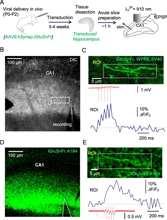
Monitoring glutamate release from multiple axons ex vivo in hippocampal slices labelled with iGluSnFR through viral transduction in vivo. a A diagram depicting viral ICV injections in neonates (P0-P2) followed by AAV transduction (3–4 weeks), dissection of hippocampi, and acute slice preparation for two-photon excitation imaging coupled with electrophysiology (Schaffer collateral stimulation and fEPSP recording in S. radiatum). b Experimental arrangement as seen in the microscope (DIC channel); stimulating and recording electrodes are seen; dotted rectangle, ROI for imaging. c Image, axon fragment in S. radiatum (ROI as in B) as seen in the green channel (AAV9.hSynap.iGluSnFR.WPRE.SV40 fluorescence; 50-frame average). Upper trace, fEPSP response to afferent stimuli (five at 20 Hz, one-trial example); lower trace, the corresponding ROI-averaged ΔF/F0 signal time course (one-trial example). d Arrangement as in (b) but for the ‘slow-decay’ sensor variant AAV2/1.hSyn.SF.iGluSnFR.A184S (green channel shown). e Experiment as in (c) but for AAV2/1.hSyn.SF.iGluSnFR.A184S; notation as in (c)

