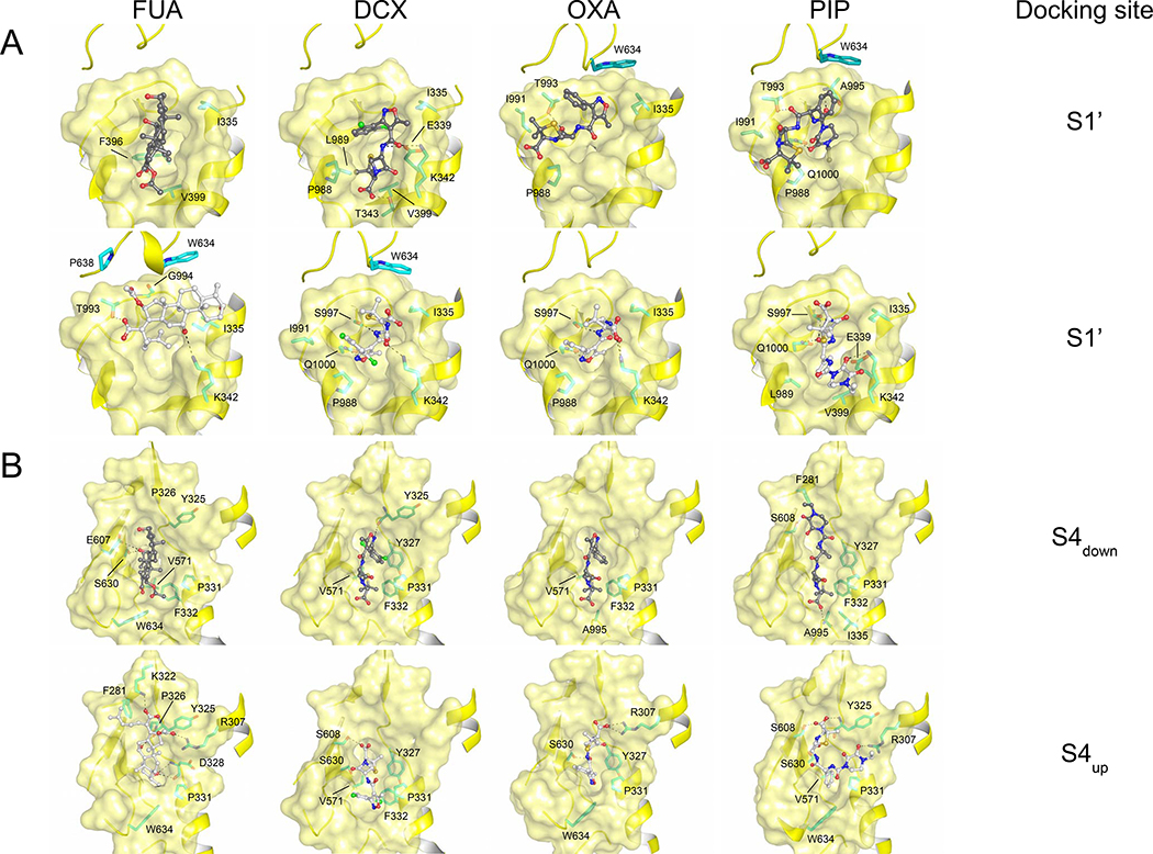
Figure 4. Potential binding conformations of FUA and carboxylated β-lactams at the S1’ and S4 docking site.
(A) S1’ binding site (upper part: carboxyl moiety of drug molecules is oriented towards TM; lower part: carboxyl moiety of drug molecules is oriented away from TM), and (B) S4 binding site (S4down: carboxyl moiety of drug molecules is oriented towards S1’; S4up: carboxyl moiety of drug molecules is oriented towards upper part of the cleft between PC1 and PN2 subdomain).Cartoon and surface representation are colored in yellow. Drug molecules are depicted as ball and stick (carbon = black or white; oxygen = red; nitrogen = blue; chlorine = green). Residues are depicted as stick models (carbon = green).

