Fig. 5.

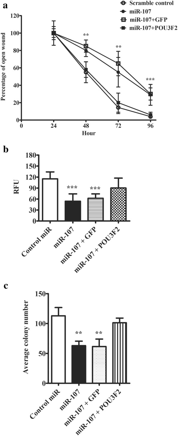
Over-expression of POU3F2 antagonized miR-107 inhibitory effects on melanoma cells. a Graphic summary of cell migration rate in wound closure experiment. SH-4 cells were co-transfected with miRNAs and POU3F2 or GFP. An open wound was created in the centre of a confluent monolayer of transfected cells. The percentage of the remaining open wound was recorded every 24 h for 4 days. b Cell invasive potential of transfected cells was measured with CytoSelect™ 24-Well Cell Invasion assay with type I collagen based matrix. SH-4 cells were co-transfected with miRNAs and POU3F2 or GFP. The transfected cells were plated on the upper chamber of a tran-well system. 48 h later, the matrix was collected and the invaded cells were stained with CyQuant® GR dye and quantified in a fluorescence plate reader. c Colony formation study of SH-4 cells co-transfected with miRNAs and POU3F2 or GFP. The colony formation ability of the transfected cells was accessed by counting the number of colonies (more than 50 cells) in the culturing plates. The data were presented as mean ± SD of at least three independent experiments (*p < 0.05, **p < 0.01, ***p < 0.001 as compared with control)
