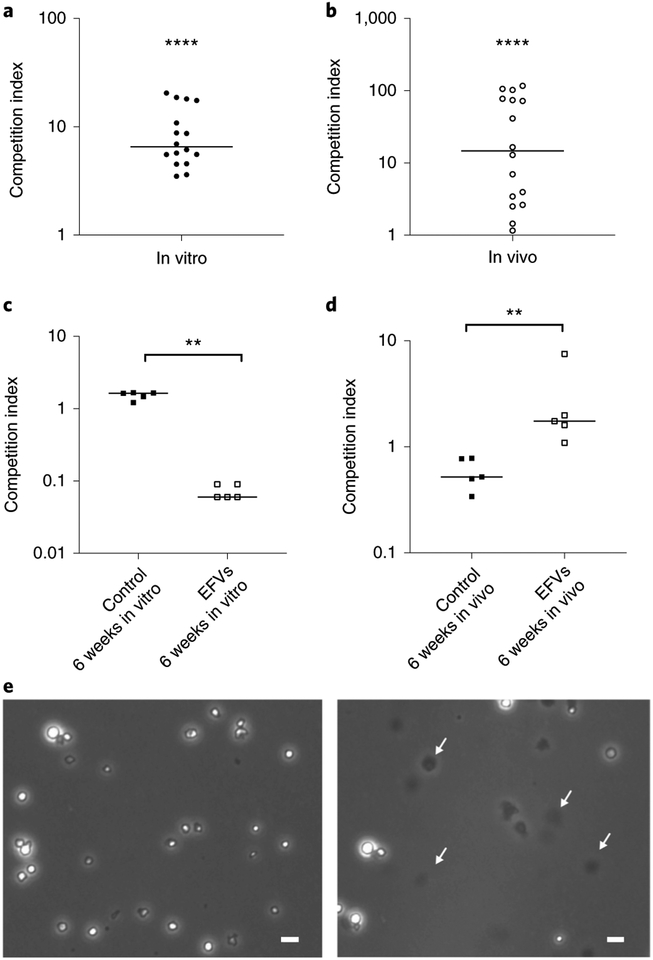
Fig. 6 |. Competition index for in vitro and in vivo assays of V. cholerae EFVs versus planktonic V. cholerae and working model.
a,b, Competition index of in vitro (a) and in vivo (b) assays calculated by the output ratio after incubation (in vitro: overnight, 37°C; in vivo: 24 h, 24°C) corrected by the input ratio. Number of colony-forming units was assessed by plating on LB10 agar plates supplemented with 100 μg ml−1 rifampicin to inhibit other intestinal bacteria and 80 μg ml−1 X-gal. Data in a and b are from 16 independent biological replicates and are shown as the median. The competition index of V. cholerae EFVs compared to planktonic cells in in vitro and in vivo assays is significantly higher than a hypothetical median of 1.0 (two-tailed, non-parametric, Wilcoxon signed-rank test; ****P< 0.0001). c,d, Competition index of in vitro (c) and in vivo (d) assays performed with either six-week-old EFVs (incubated in 0.55× NSS at room temperature) or six-week-old planktonic cells (control, incubated in 0.55× NSS at room temperature) compared with ΔlacZ wild type, calculated by the output ratio after incubation (in vitro: overnight, 37°C; in vivo: 24 h, 24°C) corrected by the input ratio. Colony-forming units were assessed by plating on LB10 agar plates supplemented with 100 μg ml−1 rifampicin to inhibit other intestinal bacteria and 80 μg ml−1 X-Gal. Data in c and d are from five independent biological replicates and are shown as the median. The competition index of the six-week-old V. cholerae EFVs compared with the six-week-old planktonic cells (control) was significantly different in both in vitro and in vivo conditions (two-tailed, non-parametric Mann–Whitney test; **P<0.01). e, V. cholerae EFVs incubated at 37°C for 4h in 0.55× NSS at pH 3.4 without (left) or with (right) 0.4% deoxycholic acid treatment. Images show intact V. cholerae EFVs in the untreated condition (left), with arrows showing digested EFVs after deoxycholic acid treatment. Scale bars, 10 μm. Images are representative of three independent experiments.

