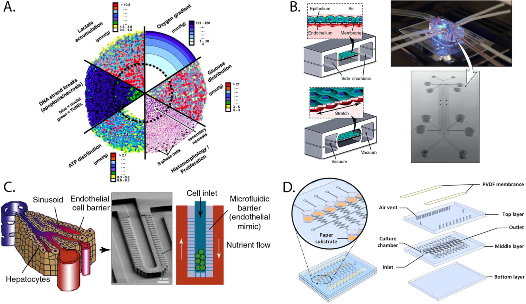
Figure 1.

Various preparations of 3D cultures. (A) Compilation of images displaying the gradients of proliferation, viability, oxygen, and nutrients that form across spheroids. Part A was reproduced with permission from ref 12. Copyright 2010 Elsevier. (B) Schematic of a lung-on-a-chip model containing a coculture of human alveolar epithelial cells and pulmonary capillary endothelial cells on the opposite sides of a membrane (left), image of the assembled device (top right), and scanning electron micrograph of the device before assembly (bottom right). (C) A liver-on-a-chip device that uses a series of microfabricated posts to mimic the endothelial barrier in the liver sinusoid, separating hepatocytes from a surrounding flow channel. Parts B and C were adapted with permission from ref 30. Copyright 2011 Elsevier. (D) Three-layer PDMS/paper hybrid microfluidic device used for uropathogen testing. The orange areas (left) contain the cellular culture chambers in paper. Part E was reproduced with permission from ref 97. Copyright 2016 American Chemical Society.
