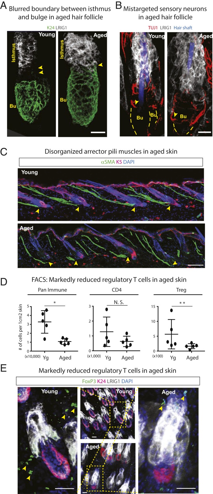
Fig. 4.
Multifaceted changes in the aged skin microenvironment. (A and B) Whole-mount immunofluorescence of telogen-phase young and aged HFs immunolabeled for HFSC marker K24, isthmus marker LRIG1, and sensory neuronal marker TUJ1. Arrowheads in A point to the spatial separation between the bulge (Bu) and isthmus. In young HFs, this zone harbors the four sensory neurons that wrap around the HFs. In aged HFs, this zone is missing. Arrowheads in B show that sensory neurons have relocalized to the bulge in aged HFs. (Scale bars, 20 μm.) (C) Immunofluorescence of α-smooth muscle actin (αSMA) in young and aged skin reveals that APMs lose their anchorage (arrowheads) to the bulge in aged skin. (Scale bar, 50 μm.) (D) FACS analysis of skin resident immune cell populations showing age-related reductions in CD45+ pan-immune populations, no significant change in the overall cohort of CD4+ effector T cells, but specific reductions in CD4+FOXP3+CD25+ Tregs. n = 5. Data are presented as mean ± SEM. Paired t test was performed, *P < 0.05, **P < 0.01, N.S., not significant. (E) Whole-mount immunofluorescence of telogen-phase young and aged HFs immunolabeled for HFSC marker K24, isthmus marker LRIG1, and Treg marker FOXP3. Arrowheads in E point to the FoxP3+ Treg cells in the young and aged skin. (Scale bars, 30 μm.) For A–C and E, at least five independent biological replicates were analyzed. Shown are representative images.

