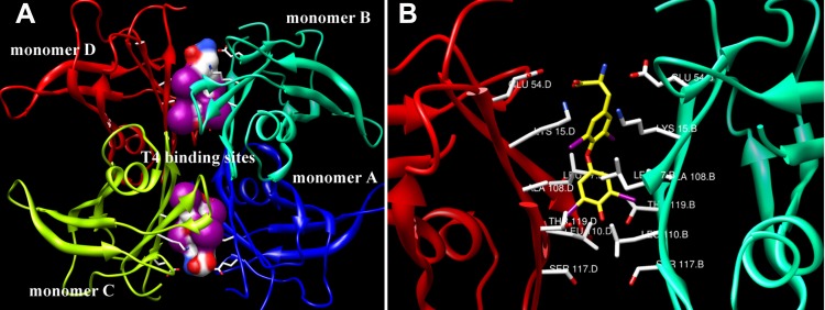
Figure 1.
Crystal structure (PDB ID: 1IE4) of TTR tetramer with T4 interacting with the two T4-binding sits is shown. (A) The two T4-binding sites located at the dimer–dimer interfaces are framed by the white boxes. (B) The specific interaction between T4 and amino acids at the binding pockets is shown. The yellow rod structure is indicated as T4, and the green solid lines are hydrogen bonds. These pictures are prepared using the program UCSF Chimera developed by the University of California.

