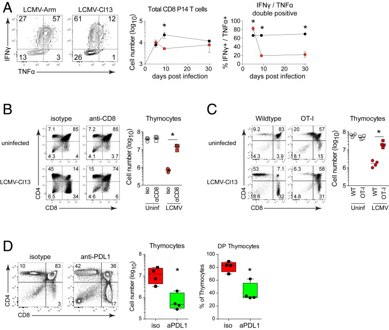
Fig. 3.
Virus-specific CD8 T cells mediate thymic depletion and their exhaustion in chronic infection enables reconstitution. (A) Mice received LCMV-specific P14 CD8 T cells and were infected with LCMV-Arm or LCMV-Cl13. Flow plots show IFNγ and TNFα expression following LCMV-GP33 peptide stimulation of thymus derived P14 cells. Graphs indicate the number of thymus-infiltrating and the percent of cytokine producing P14 T cells in the thymus on the indicated day following LCMV-Arm (black) or LCMV-Cl13 (red) infection. (B) WT mice were treated with isotype control or anti-CD8 depleting antibody and then either remained uninfected or were infected with LCMV-Cl13 infection 1 day later. Flow plots show thymus depletion and the graph demonstrates total thymic cellularity 9 d after antibody treatment. (C) Thymus depletion and cellularity in uninfected and LCMV-Cl13 infected (day 9) WT and OT-1 transgenic mice. (D) WT mice were treated with isotype control or anti-PDL1 antibody beginning 25 d after LCMV-Cl13 infection. Flow plots show thymocyte profile and graphs indicate total cellularity and the percent of double positive thymocytes. Data are representative of two to three independent experiments with four to five mice per group. Error bars indicate SD. *P < 0.05.

