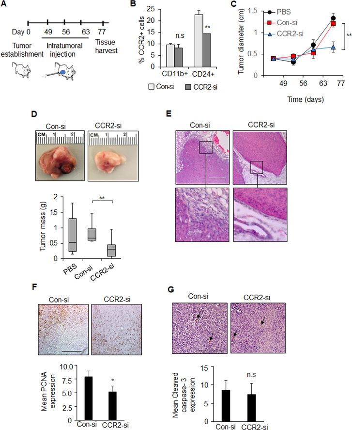
Figure 1. Effect of CCR2 siRNA delivery on PyVmT mammary carcinoma growth and invasion.
A. Experimental design: PyVmT mammary tumors were established though mammary intraductal injection of FVB mice. When tumors reached 0.4 cm in diameter, mice received intratumoral injections of PBS vehicle control, 10 μg TAT peptides complexed to control siRNAs (Con-si) or CCR2 siRNAs (CCR2-si), n=7 per group. Mice were sacrificed when control tumors reached approximately 1.5 cm in diameter, 77 days after cellular transplantation. B. Flow cytometry analysis was conducted on tumor cell suspensions for percentage of CCR2+ cells in mammary epithelial cells (CD24+) vs. myeloid cells (Cd11b+). C. Tumor diameter was measured over time by caliper. D. Mean tumor mass at endpoint. Representative tumor tissues shown. E-G. Mammary tumors were stained by H&E (E), or immunostained for PCNA expression (F) or cleaved caspase-3 (G). Arrowheads depict positive staining. Scale bar=200 microns. Staining was quantified by Image J. Expression was normalized to hematoxylin staining. Statistical analysis was determined by Two Tailed T-test (B,E,F) or One Way ANOVA with Bonferonni post-hoc analysis (C,D). Statistical significance was determined by p<0.05. *p<0.05, **p<0.01, ns=not significant. Mean+SEM are shown.

