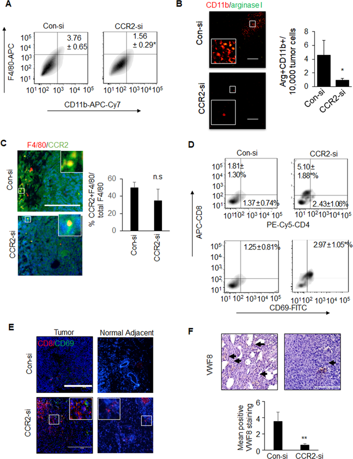
Figure 2. CCR2-deficient PyVmT mammary tumors exhibit alterations in immune cell activity and decreased angiogenesis.
PyVmT mammary tumors treated with Con-si or CCR2-si were analyzed for A. F4/80+CD11b+ macrophages by flow cytometry B. M2 macrophages by co-immunofluorescence staining for arginase I (green) and CD11b (red), C. co-immunofluorescence staining for CCR2 (green) F4/80 (red) expression, D. flow cytometry analysis for CD4+CD8+ or CD8+CD69+ lymphocytes, E activated cytotoxic T cells by co-immunofluorescence staining for CD8 (red) and CD69 (green) expression, F. angiogenesis by immunostaining for Von Willibrand Factor 8 (VWF8), arbitrary units are shown. Black arrows indicate positive staining. N=6 tumors per group, with >15 images per tumor section. Immunostaining was quantified by Image J, and normalized to DAPI (B, C, E) or hematoxylin (F). Arbitrary units are shown Statistical analysis was performed using Two-tailed T test.. Statistical significance was determined by p<0.05. *p<0.05. Mean±SEM are shown. Scale bar= 100 microns.

