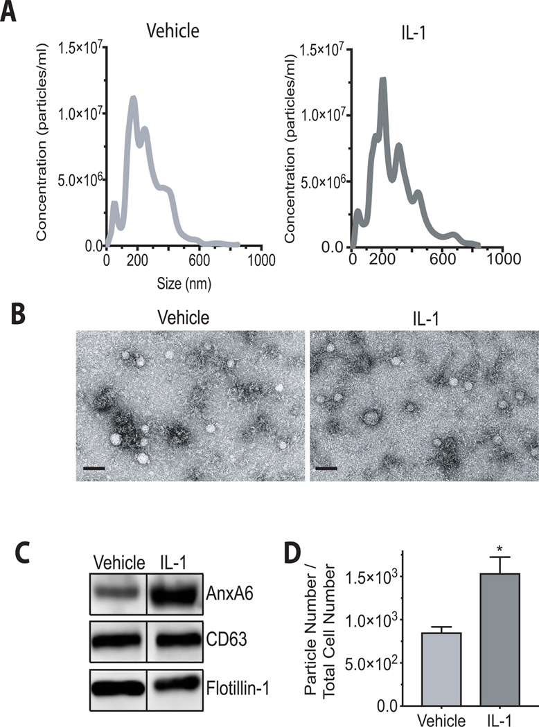
Figure 2: Characterization of EVs released from vehicle-treated and IL-1β-treated human articular chondrocytes.
EVs released into the medium from human articular chondrocytes in the absence or presence of IL-1β. Human articular chondrocytes were serum starved for 24h followed by treatment with IL-1β (10ng/ml) in PBS/0.1%BSA (IL-1) or PBS/0.1%PBS (Vehicle) for 24h. A: Size distribution of EVs in the medium of vehicle-treated (Vehicle) and IL-1β-treated (IL-1) human articular chondrocytes was measured using nanoparticle tracking analysis. B: Representative transmission electron microscopy images of EVs isolated from the medium of vehicle-treated (Vehicle) and Il-1b-treated human articular chondrocytes. Bar, 200 nm. C: Immunoblot analysis of EV markers, AnxA6, CD63, and flotillin-1 in isolated EVs from the medium of vehicle-treated (Vehicle) and IL-1β-treated (IL-1) human articular chondrocytes. D: The numbers of EVs released into the medium of IL-1β-treated or vehicle-treated cells were analyzed by nanoparticle tracking analysis and normalized to the total cell number. Data were obtained from four different experiments and expressed as mean ± SD. *p < 0.01 vs. vehicle-treated cells.

