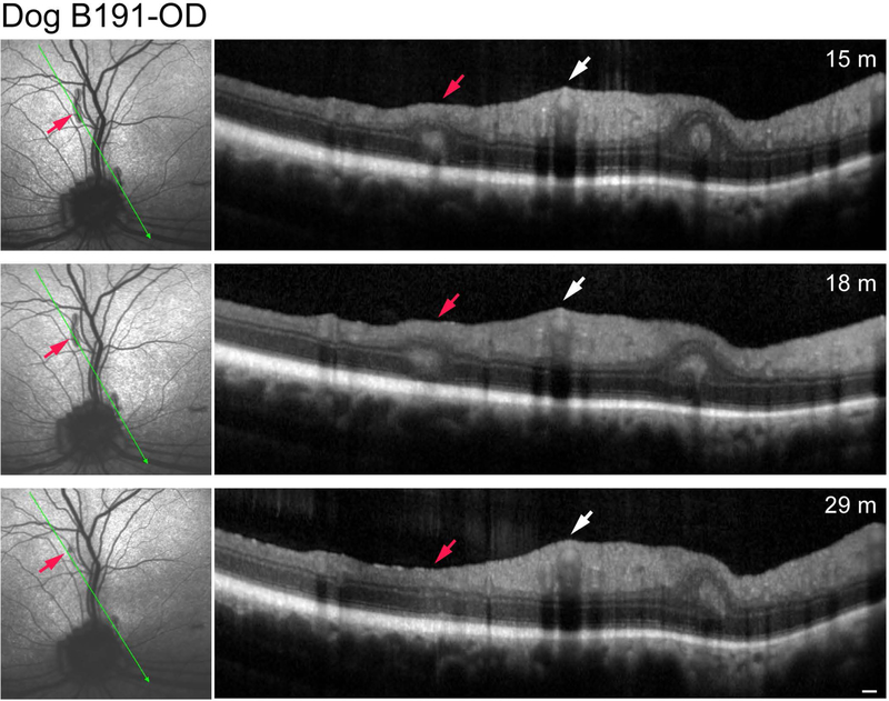
Figure 2.
Serial sdOCT scans of the same retinal fold of Dog B191-OD; plane of scan noted by thin green arrow. The red arrow points to the retinal fold that partially disappeared by 29 months of age. White arrow points to the normal thickness of inner retina at the site of the superior venule and arterioles. Note that the small fold present adjacent to and nasal to the optic disc remains unchanged. Scale bar = 200 μm for OCT scans.

