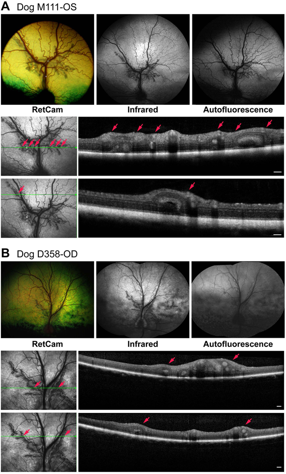
Figure 3.
Clinical and imaging findings in dogs with multifocal retinal dysplasia and having a heterozygous mutation in drd1 (A, Dog M111) or drd2 (B, Dog D358). In both cases the folds cluster perivascularly mainly along the superior venules and arterioles. For each group of images, the lower set represent the sdOCT scans illustrated for the plane of the horizontal green arrow. Small red arrows identify the specific folds identified in the en face image and OCT scans. Scale bar = 200 μm for OCT scans.

