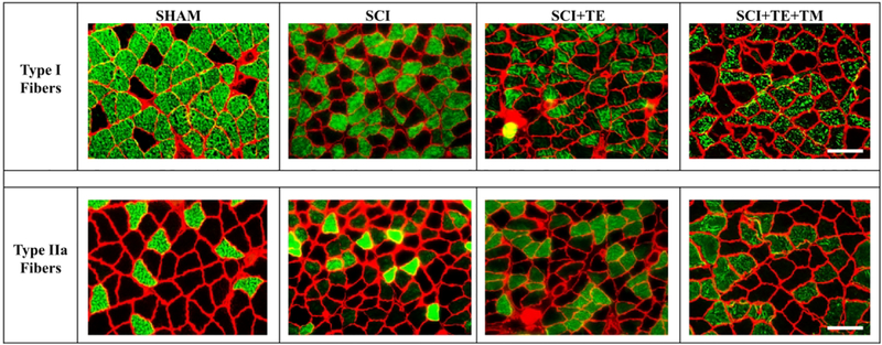
Figure 6.
Histological images of soleus type I (top row) and type IIa (bottom row) fiber-type staining from animals in our primary study after sham surgery (T9 laminectomy) or spinal cord injury (SCI) alone or in combination with testosterone-enanthate (SCI+TE) or TE plus bodyweight-supported treadmill training (SCI+TE+TM). Samples were stained with anti-MHC-I (green fibers – top row) or anti-MHC IIA (green fibers – bottom row), as described in the methods and according to our published methods (34). Scale bar = 50 μm.

