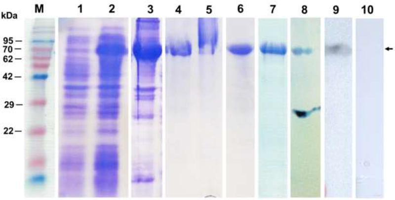
Fig. 2. SDS-PAGE and Western blot analysis of samples from expression, purification and refolding of insoluble rOv-TSP-2- LAP protein.
Protein marker (lane M), pre-induction cell lysate (lane 1), postinduction cell lysate (lane 2), insoluble protein in cell pellet post-sonication (lane 3), denatured and IMACpurified protein in 6M guanidine (lane 4), column flow-through (lane 5), insoluble eluted protein (lane 6), soluble purified protein (rOv-TSP-2-LAP) in 20 mM HEPES after refolding (lane 7), western blot of rOv-TSP-2-LAP probed with anti-6 his tag serum (lane 8), western blot of rOv-TSP-2-LAP probed with hamster anti-rOv-LEL-TSP-2 serum (lane 9), western blot of rOv-TSP-2-LAP probed with hamster naïve serum control (lane 10). Molecular weight of rOv-LEL-TSP-2 protein is approximately 75 kDa.

