
Figure 1. Cell-Surface Biotinylation of Olfactory Projection Neurons in Intact Brains.
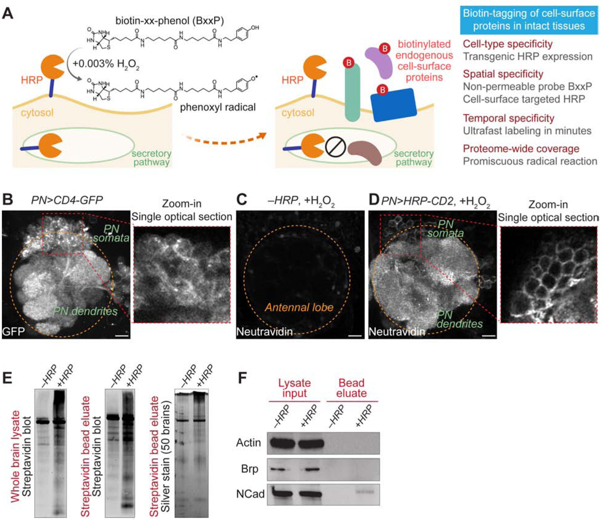
(A) Scheme and features of cell-surface biotinylation in intact tissues.
(B) Olfactory projection neuron (PN) specific VT033006-GAL4 (PN-GAL4, hereafter) drove the expression of membrane-targeted GFP (CD4-GFP). The zoom-in panel shows a single optical section of the PN soma area. Orange circle, antennal lobe.
(C and D) Neutravidin staining of antennal lobes after the cell-surface biotinylation reaction. (C) HRP was not expressed by omitting the GAL4 driver. (D) PN-GAL4 drove the expression of cell surface-targeted HRP (HRP-CD2). The zoom-in panel shows a single optical section of the PN soma area.
(E) Left and middle, streptavidin blots of the whole-brain lysate (left) and the post-enrichment bead eluate (middle). Right, silver stain of the post-enrichment bead eluate. −HRP, PN-GAL4 omitted; +HRP, PN>HRP-CD2.
(F) Immunoblots of intracellular proteins, actin and bruchpilot (Brp), and neuronal surface protein N-cadherin (NCad) in the whole-brain lysate and the post-enrichment bead eluate. −HRP, PN-GAL4 omitted; +HRP, PN>HRP-CD2.
Scale bar, 10 μm.
See also Figure S1.