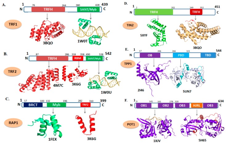
Figure 2.
Domain organization and tertiary structure of the shelterin complex. (A) telomere repeat factor 1 (TRF1) (PDB ID: 3BQO; depicts the cartoon representation of TRF homology (TRFH) domain of TRF1 in red. 1W0T; the DNA-binding domain of TRF1 shown in green in complex with telomeric DNA which is shown as light orange). (B) TRF2 (PDB ID: 4M7C; represents TRFH domain of TRF2. 3K6G; represents the repressor/activator protein 1 (RAP1)–binding motif of TRF2 in red. 1W0U; represents the DNA-binding motif of TRF2). (C) RAP1 (PDB ID: 1FEX; represent the Myb domain shown in green and 3K6G; shows RCT domain in red). (D) TIN2 (PDB ID: 5XYF; cartoon representation in green depicts TRFH domain and 3BQO; TRF1-interacting nuclear factor 2 (TIN2) peptide shown in red complexed with TRFH domain of TRF1). (E) TPP1 (PDB ID: 2I46; depicts the OB-fold domain in purple and 5I2X; represents the POT1 interacting domain of TPP1 (cyan) with C-terminal domain of POT1 (gray)). (F) POT1 (PDB ID: 1XJV; depicts the C-terminal domain of POT1 in purple in complex with telomeric DNA (light orange) 5H65; represents the POT1 interacting domain of TPP1 (red) with C-terminal domain of POT1 (purple).

