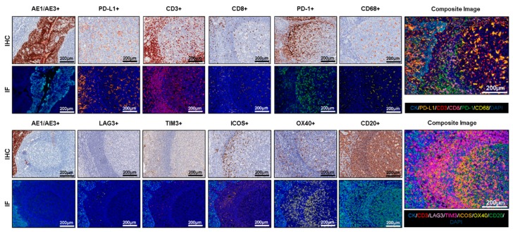
Figure 2.
Antibody optimization and validation. Representative examples of antibody optimization and validation using conventional chromogenic IHC and multiplex IF showing similar patterns of expression by the antibodies tested in IHC and multiplex IF. Composite images show the integration of markers on a single slide. At 20× magnification.

