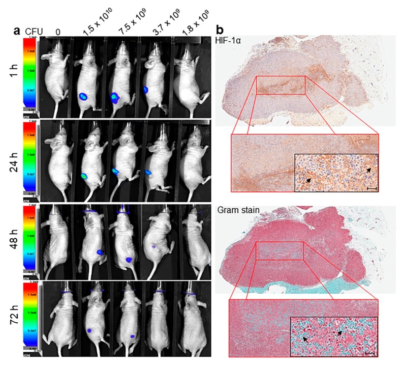
Figure 4.
Detection of L. lactis-IRFP713 via advance molecular imaging and co-localization of L. lactis with HIF-1α in the core of the melanoma tumor. (a) Representative time-course imaging of tumor bearing mice administered with IRFP713-expressing L. lactis. BALB/c mice were administered 1 × 10⁶ A375 cells in the right flank by s.c. injection. Ten days after tumor inoculation, mice were administered IRFP713-expressing L. lactis at increasing concentrations (0, 1.8 × 10⁹, 3.7 × 10⁹, 7.5 × 10⁹ and 1.5 × 10¹⁰ CFU) by i.t. injection. Whole body imaging was performed at 1, 24, 48 and 72 h with the AMI-1000-X instrument under 690/713 nanometers (nm) excitation and emission visualization, respectively; (b) Representative IHC staining of HIF-1α and Gram staining of A375 tumor sections from BALB/c mice injected i.t. with L. lactis-IRFP713 at a magnification of ×100 and ×400. Arrows indicate gram-positive bacteria cells and cells positive for HIF-1α, respectively. Whole tissue slides were scanned with Leica Aperio ImageScope with 40× magnification. Scale bar = 50 µm.

