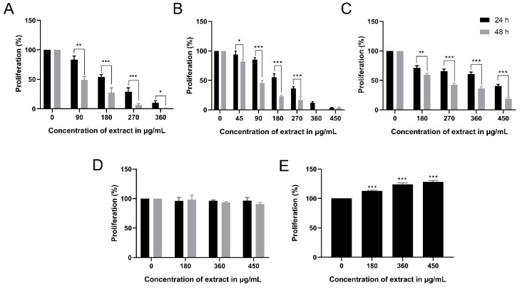
Figure 1.
Proliferation of Monomac-1 (A), KG-1 (B), U937 (C), mesenchymal stem cells (MSCs) (D), and normal mononuclear cells MNCs (E), after 24 h and 48 h of treatment with methanolic leaf extract (MMLE). A significant dose- and time-dependent inhibition of proliferation of the three AML cell lines was noticed with increasing concentrations of MMLE. Significant differences were reported with * indicating a p-value: 0.01 < p < 0.05, ** indicating a p-value: 0.001 < p < 0.01 and *** indicating a p-value: 0.0001 < p < 0.001.

