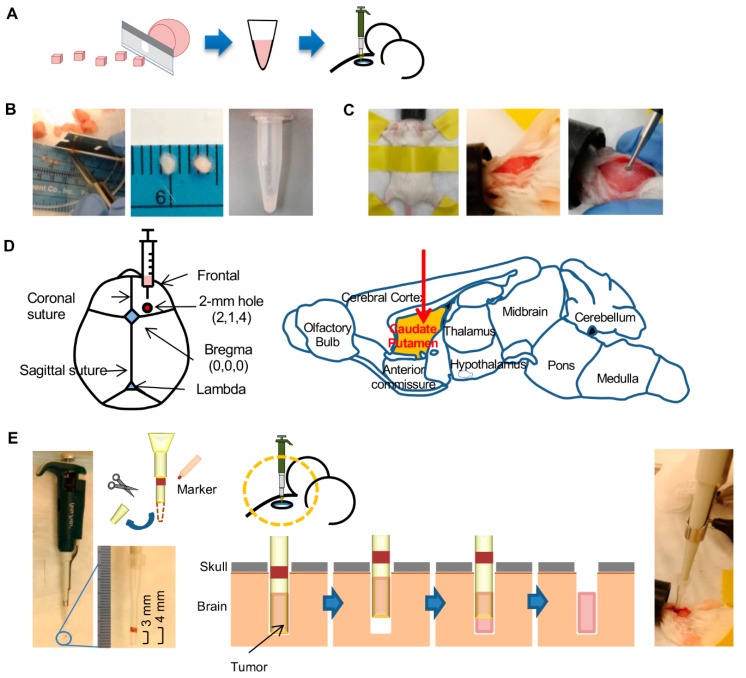
Figure 1.
Method for implantation of tumor in mouse brain with the pipette method. (A) Workflow of the method involves mincing the tumor into ~1 mm3 pieces with a blade. Size of the pieces is then further reduced by crushing the pieces in medium (2× volume) with a pipettor tip before injection of the preparation into mouse brain. (B) Photographs shows mincing of a tumor, some minced tumor pieces (ruler markings are in mm), and the injection-ready tumor preparation after crushing of the pieces with a pipette tip. (C) Preparation of mouse for tumor injection. An anesthetized mouse is secured in the prone position with tape. The crown of the head is shaved, and a 10-mm incision is made along the midline of the skull. A burr hole of 2 mm width is then made 2 mm lateral and 1 mm anterior to the bregma with a micro drill. (D) Coordinates for tumor injection are depicted on anatomic maps of mouse skull and brain. The 2-mm wide burr hole is at X, Y, and Z coordinates of 2 mm, 1 mm, and 4 mm with respect to the bregma. Tumor preparation is injected in the caudate putamen of brain. (E) Method of injection of the tumor preparation. The distal 1 cm of a 10-µL pipette tip is cut off with scissors, and a 1-mm wide marking is made on the tip at 3 mm distance from the tip’s opening. Tumor preparation (3 µL) is picked up with the tip attached to a pipette (photograph on left). The tip is then placed through the burr hole (photograph on right) to a depth of 4 mm. After withdrawing the tip by 1 mm, the tumor preparation is ejected by pushing the pipette plunger. The tip is then completely withdrawn, and the burr hole is sealed with bone wax and the scalp is sutured.

