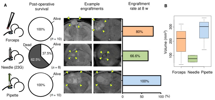
Figure 2.
Comparison of three methods for tumor implantation in mouse brain. Brain metastasis of one triple negative breast cancer patient that had been passaged three times as xenograft in mouse brain was used for this experiment. (A) Three methods were used for implantation of mouse brain with tumor. The methods used forceps and tissue blocks (n = 10), 23 G needle and minced tumor tissue (n = 8), or pipette tip and minced tumor tissue (n = 10). 1 µL volume of tumor that had been passaged three times as xenograft was used for each mouse. Pie charts depict survival rates within 1 day of implantation with the three methods. Examples of coronal sections of brain in magnetic resonance imaging are shown for two mice each for the three methods. The extent of tumors in the images are indicated with arrowheads. Rates of engraftment of implanted tumor after 8 weeks of surgery for tumor implantation are plotted for the three implantation methods. Mice that did not survive surgery are excluded. (B) Tukey boxplots of tumor volumes at 6 weeks in mice with engraftment are shown for the Forceps, Needle, and Pipette methods.

