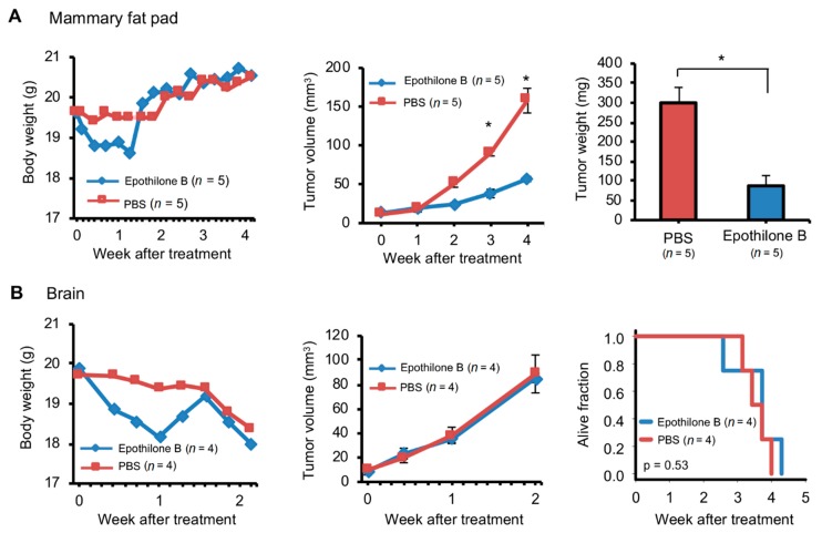
Figure 6.
Different responses to chemotherapy of breast cancer patient brain metastasis implanted in the mouse brain or mammary fat pad. Brain metastasis of one triple negative breast cancer patient was used for this experiment. Tumor was prepared with the mincing method for implantation (1 µL tumor + 2 µL phosphate-buffered saline [PBS]) in the mouse mammary fat pad (A) or brain (B) with the Pipette or Forceps method respectively. Implanted tumors were then passaged three times at the same implantation site. Mice with tumors after third passage were intravenously injected with one dose of epothilone (4 mg/kg) or PBS when tumors were ~10 mm3 in volume. Growth of tumors was measured with calipers (A) or magnetic resonance imaging (B). Body and tumor weights and tumor volumes are plotted (mean and standard error). p values in standard t tests for group comparison are indicated (* ≤ 0.05). Survival plot for mice with tumors implanted in brain is also shown (p value determined with logrank test).

